|
Imaging the
Brain
|
|
|
|
|
|
DOE scientists
from many disciplines are collaborating to develop and apply new technologies
to study the function of the human brain. The goals of the research
are to expand our basic understanding of how the brain works and to
develop tools for the diagnosis and treatment of mental and neurological
disorders.
by John George.
LANL
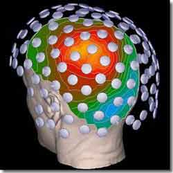
A
representation of the magnetic fields measured by sensors
as superimposed on the surface of the subject's head.
|
The approach involves advanced sensor technologies, complex system
integration, mathematical and computer modeling, and computational
visualization. At Los Alamos National Laboratory, the primary method
of noninvasive functional brain imaging being investigated is magnetoencephalography
(MEG) and the integration of MEG with magnetic resonance imaging
(MRI) and functional MRI (fMRI).
MEG measures minute magnetic fields produced by currents in electrically
active nerve cells (neurons) in the brain. These minute magnetic
fields can be measured by superconducting quantum interference device
(SQUID) sensors, the most sensitive magnetic field detectors known,
which are arranged in an array over the surface of the head. MEG
is a completely noninvasive and passive (not even using externally
applied magnetic fields) method of measuring brain function. MEG
data input to sophisticated computer models allows neuronal activity
to be located in space and with better than millisecond temporal
resolution.
We have developed a new type of MEG sensor based on the Superconducting
Imaging Surface (SIS) concept. The SIS produces an image of the
magnetic source while shielding the sensors from the more powerful
external magnetic fields, such as those produced by electric motors
and Earth itself. (The magnetic field "lines" of these external
sources can not penetrate the SIS). As a result, the SIS technology
improves the signal-to-noise ratio of MEG measurements. Prototypes
based on this idea have been successfully tested and a whole-head
sensor array "helmet" based on this concept is currently being assembled.
Imaging brain function with MEG requires a series of computational
tools for collecting and processing signals, modeling the physics
of the measurement, and building probabilistic models of neuronal
currents that account for the data. Anatomical MRI is used to noninvasively
generate an image of the anatomy that is used to define the geometry
of the head and brain, for modeling, and for visualizing regions
of brain activity. Information from PET, fMRI, and other methods
can be used to further improve the accuracy and reliability of functional
brain maps based on MEG. The functional information obtained from
PET and fMRI are supportive in that they provide a map of where
activity is occurring, while the MEG provides the when, or temporal
information, simultaneously with the where.
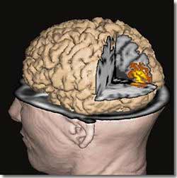
LANL
Modeling
of cortical activations during a vision experiment.
|
In spite of their tremendous value, MEG, MRI and other available
methods do not provide all of the information needed for the best
medical care. By integrating information from multiple techniques
we can exploit complementary strengths of existing methods, and
we are working with scientists who are developing technologies that
will provide new capabilities for research and clinical practice.
For example, optical tomography can provide useful information about
the biochemistry, physiology and anatomy of biological tissues,
such as the head or breast. Thus, it is possible that optical tomography
could add the what to the where and when information provided by
MEG and FMRI.
We are exploring several "spin-off" biomedical applications of
technologies developed for functional brain imaging. For example,
we are developing a method of detecting and treating cancers (recently
filed patent) and a method of detecting and locating aberrant electrical
activity in the heart (such as those that cause atrial fibrillation).
In addition, we are exploring non-biomedical applications such as
a method of detecting and characterizing defects in materials.
|
| |
|
|
|
|
|